

Extracellular vesicle characterization
Extracellular vesicle characterization uses complementary techniques to evaluate EV size, concentration, markers and physicochemical properties.
Extracellular vesicle characterization
Extracellular vesicle characterization uses complementary techniques to evaluate EV size, concentration, markers and physicochemical properties.
Extracellular vesicle characterization is the use of analytical techniques to evaluate the physical, chemical and molecular properties of extracellular vesicles (EVs), including particle size, concentration, morphology, markers, composition and surface characteristics. Because different particle populations can coexist within the same sample, complementary methods are often combined to obtain more comprehensive information about the EVs being studied.
What is extracellular vesicle characterization?
Extracellular vesicles are lipid bilayer-delimited particles naturally released by different cell types. They can carry proteins, lipids, nucleic acids and other molecules and are investigated in fields including intercellular communication, biomarker research, cancer, regenerative medicine and the development of delivery systems.
Following sample collection, preparation, isolation or enrichment, characterization helps researchers describe the particles present and evaluate their properties. A single analytical technique generally cannot provide every type of information required. EV studies therefore frequently combine physical measurements with techniques designed to investigate molecular components.
Which parameters are evaluated when characterizing EVs?
The analytical strategy depends on the scientific question and sample type. Common parameters evaluated during extracellular vesicle characterization include:
- Size: determines the distribution of particle dimensions within the sample.
- Concentration: estimates the number of particles per sample volume.
- Morphology: provides information about vesicle structure and appearance.
- Molecular markers: help investigate proteins and other molecules associated with the studied populations.
- Zeta potential: provides information related to particle surface charge and electrokinetic behavior in suspension.
- Fluorescence: can be used to investigate particles associated with specific fluorescent markers.
Techniques for extracellular vesicle characterization
Different analytical technologies can be used as complementary approaches. Nanoparticle tracking analysis (NTA) tracks individual particles undergoing Brownian motion and uses this information to determine particle size distribution and concentration. Microscopy techniques can provide morphological information, while biochemical and immunological methods can be used to investigate proteins and other molecular markers.
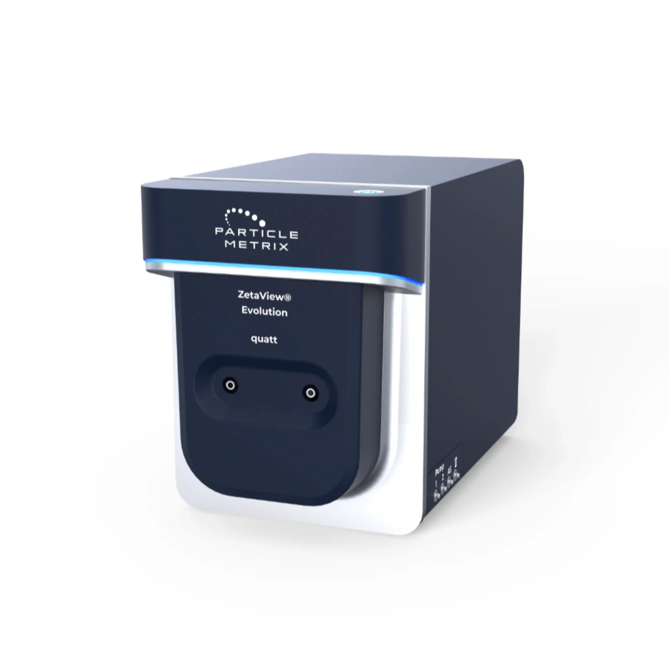
For studies that need to relate physical characteristics to fluorescently labeled populations, multiparametric platforms can expand the information obtained from particle analysis. The ZetaView Evolution supports NTA, fluorescence analysis and zeta potential measurements. Configurations with multiple lasers can also support the investigation of labeled particle subpopulations, making the technology particularly relevant to extracellular vesicle research.
Characterization of extracellular vesicle subpopulations
EV samples can be highly heterogeneous. Particles with similar dimensions do not necessarily share the same biological origin or molecular composition. Measuring size and concentration alone may therefore be insufficient when the objective is to distinguish specific populations.
Fluorescent particle analysis can add marker-specific information to physical characterization. Depending on the technology, researchers can investigate fluorescence signals associated with individual particles and study different subpopulations within a preparation. This can be especially useful for research involving surface markers and specific EV populations.
Applications of extracellular vesicle characterization
EV characterization is used in basic research, translational studies and biotechnology development. Analytical parameters should be selected according to sample origin, preparation method and experimental objectives.
- Characterization of exosomes and other EV populations.
- Biomarker discovery and validation.
- Studies of intercellular communication.
- Comparison of EV isolation and purification methods.
- Evaluation of extracellular vesicle preparations.
- Research in cancer, disease and regenerative medicine.
- Investigation of subpopulations using fluorescent markers.
What is the best technique for extracellular vesicle characterization?
No single technique can provide all the information required to characterize an EV population. The appropriate approach depends on the experimental question. Particle size and concentration measurements can be combined with microscopy, marker analysis, fluorescence and other methods to provide more comprehensive characterization.
Why are size and concentration not sufficient to identify exosomes?
Different extracellular vesicles and non-vesicular particles can have overlapping size distributions. Therefore, a particle cannot be classified as an exosome based only on its dimensions. Additional information about origin, composition and molecular markers is required for a more specific interpretation.
How does fluorescence help characterize extracellular vesicles?
Fluorescence can be used to investigate particles associated with specific fluorescent molecules or markers. When combined with individual nanoparticle analysis, it can help distinguish subpopulations within heterogeneous samples and relate physical particle characteristics to the presence of the markers being investigated.
Other terms with the letter "E"
View allExtracellular Vesicles
Extracellular vesicles are cell-released nanoparticles that carry proteins, lipids and genetic material, enabling cell-to-cell communication.
Endosomal Compartment of Cells
A system of organelles that receives, sorts, and directs internalized cellular cargo toward recycling, degradation, or other destinations.
Exosomes
Exosomes are small extracellular vesicles released by cells that transport biomolecules and participate in intercellular communication.
Extracellular vesicle isolation
Extracellular vesicle isolation separates and enriches EVs from biological fluids or culture media for subsequent analysis and characterization.
Exosome biomarkers
Exosome biomarkers are vesicle-associated molecules used to investigate EV origin, composition and potential biological functions.
Exosome purification
Exosome purification reduces contaminants and enriches vesicles of interest for subsequent physical, molecular and functional analysis.
Stay in the loop
Get updates, articles and news by email.
Expect news about equipment, services and regulations tailored to your locale.