GAVE 2026 highlights: extracellular vesicles from placental biology to vaccines
The fourth GAVE workshop brought researchers together in Buenos Aires to discuss the roles and applications of extracellular vesicles. Reliable isolation and characterization emerged as challenges across the studies presented.
GAVE 2026 highlights: extracellular vesicles from placental biology to vaccines
29 Sep 2026 14 min read
The fourth GAVE workshop brought researchers together in Buenos Aires to discuss the roles and applications of extracellular vesicles. Reliable isolation and characterization emerged as challenges across the studies presented.
From alterations in communication between placenta and endothelium to the use of bacterial vesicles as vaccine candidates, the GAVE 2026 presentations showed how extracellular vesicle research has moved toward functional questions. The studies investigated what these particles do, how they can be modified, and to what extent isolation and characterization methods support the conclusions.
In this article, we bring together the main results and debates presented at the meeting:
The role of EVs in maternal-fetal health and fertility;
Applications of bacterial vesicles in immunomodulation and vaccines;
Studies in cancer, neuroscience, and regenerative medicine;
Biomarker research in different diseases;
The challenges of EV isolation, quantification, and characterization.
About GAVE 2026
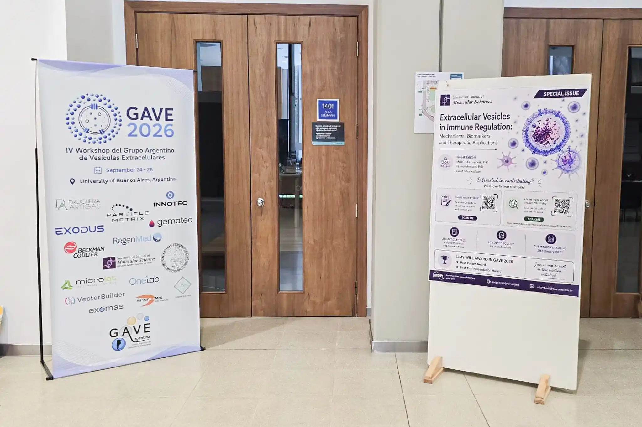
GAVE 2026, the IV Workshop of the
Argentine Group of Extracellular Vesicles
, brought together researchers on September 24 and 25, at the Cero+Infinito building of the
FCEN-UBA
, in Buenos Aires. The program covered placental biology, reproduction, microbiology, cancer, neuroscience, and regenerative medicine.
A community that grew over four editions
Created in 2022 to bring together Argentine groups dedicated to extracellular vesicles (EVs), GAVE also sought to promote international exchange. In the fourth edition, work with affiliations in Chile, Belgium, and Brazil shared space with research from Argentine institutions and the
CONICET
network.
The organizing committee brought together Ana Colado (
SphereBio
), Luis Exequiel Ibarra (
INBIAS
), Paula Soledad Pérez (
INBIRS
), Carolina Poncini (
IMPaM
), Gabriel Paparini and Antonella Filizzola (
IQUIBICEN
). The diversity of the program reflected the composition of the field itself: different organisms, biological matrices, and experimental questions require analytical approaches that are not always interchangeable.
Maternal-fetal health: vesicles in placental communication
Hypoxia-reoxygenation and endothelial migration
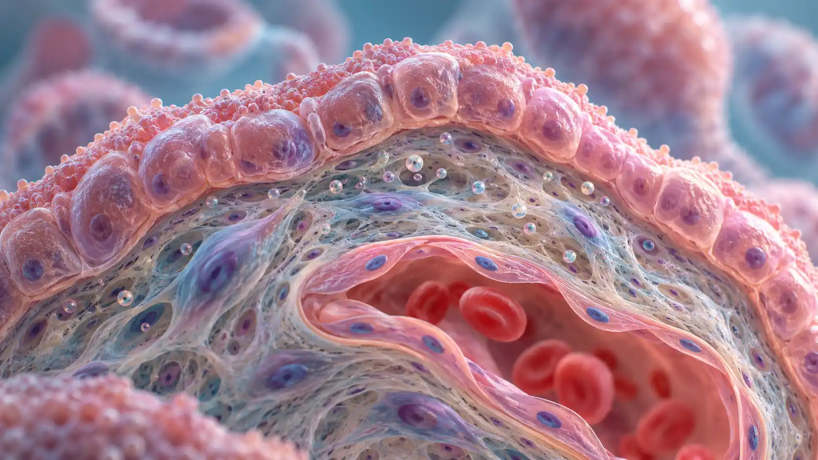
Placental biology formed one of the densest axes of the workshop. In a study by the Laboratory of Biomembranes “Prof. Dr. Mario Parisi” (
IFIBIO Houssay
, UBA-CONICET) with the
CEFyBO
, trophoblast-derived EVs produced different effects on the migration of placental microvascular endothelial cells depending on the condition of origin.
EVs obtained under normoxia favored migration. Those produced under hypoxia-reoxygenation impaired the process; treatment with melatonin partially reversed that effect. The model is relevant for investigating communication between trophoblast and endothelium under conditions associated with compromised placental perfusion, such as preeclampsia. The functional result shows why knowing only the quantity of released EVs is not enough to interpret their role.
Anandamide and myometrial activation
Another study from CEFyBO examined anandamide in the release of placental EVs and evaluated the effect of these vesicles on myometrial activation. The proposal is to investigate a possible axis between anandamide, placental vesicles, and myometrium at the onset of uterine activity.
Infection, autophagy, and trophoblast function
The program also addressed the interaction between infection and placenta. Researchers from IQUIBICEN and
IBYME
studied outer membrane vesicles of
Porphyromonas gingivalis
, a bacterium associated with periodontitis. The vesicles were internalized by trophoblast cells and compromised autophagic flux, favoring intracellular bacterial persistence. In the model presented, rapamycin partially restored trophoblast migration.
Fetal sex and maternal immune response
A fourth study, conducted by IQUIBICEN with
IMEX
and the
Hospital Italiano de Buenos Aires
, examined a variable rarely discussed in the characterization of placental EVs: fetal sex. Vesicles from pregnancies with male and female fetuses had similar size and concentration but differed in molecular composition. They also induced distinct activation programs in circulating maternal monocytes, including differences in lipid metabolism and efferocytosis. In this case, similar physical measurements did not mean biological equivalence.
Reproduction and fertility: the origin and cargo of EVs matter
Two studies explored applications of vesicles in animal and human reproduction.
Loading SPINK-3 into seminal EVs
Researchers from the
Institute of Biological Research
(CONICET-UNMdP) and
INTA Balcarce
artificially loaded ram seminal plasma EVs with the recombinant decapacitation factor SPINK-3. Comparison between incubation at room temperature and freeze-thaw cycles showed that the method alters cargo localization. The cycles favored luminal incorporation, protected from proteolysis; incubation produced predominantly surface association. For those developing vesicles as vehicles, this difference changes the interpretation of stability and availability of the transported molecule.
Follicular fluid EVs and sperm capacitation
In another study, CEFyBO characterized EVs from human follicular fluid and investigated their effect on sperm capacitation. The vesicles were taken up by spermatozoa and reversed the effect of pharmacological inhibition of the MRP4 transporter on hyperactivation. There was no alteration of the induced acrosomal reaction, which delimits the observed effect rather than attributing a general change in sperm function to the EVs.
Bacterial vesicles: immunomodulation and vaccine development
Bacterial outer membrane vesicles, or OMVs, appeared in studies with different objectives. Some investigated their ability to induce protection; others examined interactions with human and plant cells.
Brucellosis
A consortium involving the
Leloir Institute Foundation
,
IDEHU
, and
ICTAER
evaluated OMVs from
Brucella ovis
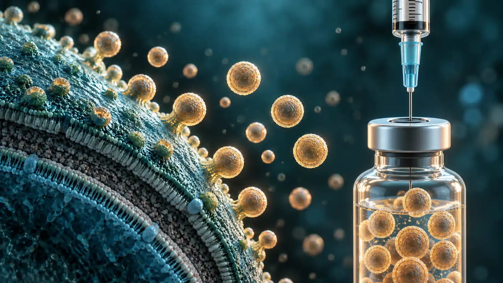
as an acellular vaccine platform for cross-protection. Vaccination in mice induced a specific humoral response and a Th1/Th17 cellular response. Sera from naturally infected pigs also recognized these OMVs, a finding that suggests antigenic conservation among species of the genus and justifies further evaluation stages.
Salmonellosis
At the
Universidad Andrés Bello
in Chile, researchers showed that high osmolarity during production modifies the physicochemical and immunogenic properties of OMVs from
Salmonella Typhi
. The condition reduced heterologous protection against
S. Typhimurium
. The study shows that, in a vesicle-based vaccine platform, culture conditions are part of the product definition.
Intestinal immunomodulation
A group from the
National University of Luján
, with the
CIC
and CONICET, observed that EVs from
Bacillus subtilis
attenuated the inflammatory response induced by LPS in human intestinal epithelial cells, without affecting cell viability.
Vesicle-mediated communication also appeared outside biomedicine. A study by UBA and
IFIBYNE
with the
VIB Center for Plant Systems Biology
in Ghent investigated OMVs from
Pseudomonas protegens
in contact with roots of
Arabidopsis thaliana
. Confocal microscopy allowed visualization of their association with the plasma membrane of root cells.
Cancer: EVs as active components of the tumor microenvironment
In cancer, two studies examined mechanisms capable of modifying communication between tumor cells and the surrounding tissue.
Imidacloprid, tumor EVs, and angiogenesis
INBIOMED
(CONICET-UBA), in collaboration with groups from Granada and the
Carlos III Health Institute
, investigated the effect of imidacloprid on HER2-positive breast cancer cells. Exposure to the insecticide increased the secretion of tumor EVs. In assays with endothelial cells, these vesicles stimulated proliferation, migration, and formation of tubular structures, results compatible with the promotion of angiogenic processes in the model studied. The work proposes a vesicular pathway for investigating the relationship between environmental exposure and tumor behavior.
Clusterin and EV release in glioblastoma
Meanwhile, INBIRS, with the
Hospital Fernández
and the
Josep Carreras Leukaemia Research Institute
, studied clusterin in gliomas. Its silencing in glioblastoma cells markedly reduced EV release. Thus, in addition to its association with tumor aggressiveness, the protein appears as a participant in vesicle production in that model.
Neuroscience: from molecular cargo to therapeutic delivery
The neuroscience studies followed three stages of the same question: identifying what EVs carry, analyzing differences in their composition, and testing whether they can deliver a cargo with a measurable effect.
Protein profile of neural stem cell EVs
IBR
(CONICET-UNR) mapped the protein profile of small EVs derived from neural stem cells. The study combined size-exclusion chromatography, MISEV-guided proteomics, and interaction network analysis. The described cargo included biogenesis markers and proteins linked to energy metabolism, antioxidant defense, and synaptic plasticity.
Single-vesicle nanoscopy in Alzheimer's disease
CIMETSA
, from the University Institute of Biomedical Sciences of Córdoba, presented results on EVs derived from human stem cells in the context of Alzheimer's disease. Among the approaches used, STED nanoscopy allowed comparison of neuroglial cargo at the level of individual vesicles between control samples and samples related to the disease.
Intranasal delivery of a therapeutic cargo
A study by the
IIB
and the
School of Bio and Nanotechnologies
(UNSAM-CONICET) tested EVs loaded with a plasmid for the neuronal glycoprotein M6a. Administered intranasally to mice subjected to chronic stress, they restored body weight gain and reduced
grooming
latency. Control EVs loaded with GFP did not reproduce these effects. The results support investigation of the strategy in the experimental model, without anticipating its clinical application.
Vesicle engineering and regenerative medicine
Modifying the surface, controlling release, and choosing the isolation method were three paths presented to bring EVs closer to therapeutic applications.
Surface engineering for regeneration
INBIRS, with the IMEX of the
National Academy of Medicine
, developed a fusion protein to anchor the Gas6 binding domain to the vesicular membrane. In mice, the modified EVs accelerated wound closure. They also reduced the ability of Langerhans cells to stimulate lymphocyte proliferation, combining regenerative and immunomodulatory effects in the model evaluated.
Controlled release in PVA nanofibers
At the
National University of Villa Mercedes
in San Luis, EVs from HEK-293T cells were incorporated into PVA nanofibers produced by electrospinning. The
scaffold
delayed vesicle release, a strategy aimed at the problem of short half-life and low retention of EVs at the lesion site. The materials preserved structural integrity and showed biocompatibility results according to the ISO 10993-5 criteria used in the study.
Quantitative recovery or morphological purity
IQUIBICEN, in collaboration with the
University of Seville
, compared ultracentrifugation and immunomagnetic separation to isolate EVs from human amniotic epithelial stem cells and from derived cells with a hepatocytic phenotype. Immunomagnetic separation favored quantitative recovery; ultracentrifugation produced preparations with greater morphological purity and uniformity. The choice of method therefore depends on the property the experiment needs to preserve or maximize.
Biomarkers: vesicular signals in different diseases
The search for biomarkers covered parasitoses, metabolic diseases, and graft evaluation. In each case, the question was similar: what information does an EV preparation add to the reading of the biological process?
Vesicular microRNAs in cysticercosis
IMPaM and
INIBIOLP
, with participation from the
Vale Technological Institute
, characterized microRNAs secreted
in vitro
by metacestodes of
Taenia crassiceps
, an experimental model for cysticercosis. The small RNAs were predominantly associated with EVs. The group also identified miRNA-binding proteins in cestodes, including AGO-2, expanding the molecular basis for investigating diagnostic candidates.
EV quantity and proteome in Chagas disease
In Chagas disease, researchers from IMPaM, the
Hospital Ramos Mejía
, and the
Oswaldo Cruz Institute
studied EVs released by peripheral blood mononuclear cells from chronic patients, grouped according to the severity of cardiac impairment. Patients in the intermediate category released more vesicles, a result interpreted as a possible sign of cell pre-activation.
In a complementary study, the group applied mass spectrometry proteomics to the EVs of these patients, with partners from
UADE
and the
Carlos Chagas Institute (Fiocruz Paraná)
in Curitiba. The protein profile distinguished the group with dilated cardiomyopathy and showed enrichment in cytoskeletal, adhesion, and contractile proteins. These are candidate signatures for clinical progression studies; their predictive usefulness still depends on validation.
Plasma EVs and glucose metabolism
Researchers from the
University of Concepción
in Chile examined plasma EVs from normoglycemic, prediabetic, and diabetic individuals. Changes in vesicle structure and function accompanied glycated hemoglobin levels. In cell assays, there were alterations in glucose uptake and lipid accumulation in muscle cells, bringing vesicle characterization closer to a concrete metabolic question.
Methodology: how to trust what was isolated and measured?
If there was a cross-cutting theme at GAVE 2026, it was the reliability of analysis. The studies showed that yield, purity, sensitivity, and preservation of function do not necessarily advance together.
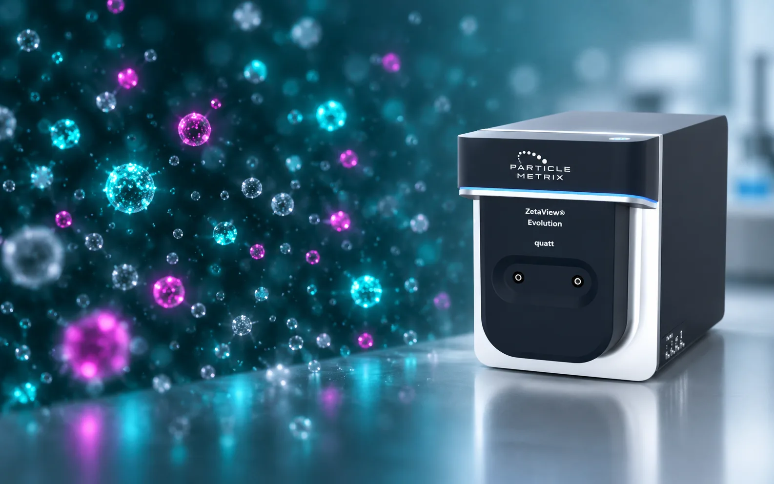
How ZetaView expands extracellular vesicle characterization
In this context, nanoparticle tracking analysis, or NTA, allows tracking the Brownian motion of individual particles in suspension. From this motion, it is possible to estimate the hydrodynamic diameter and determine the particle concentration in the sample.
The
ZetaView Evolution
brings together different analysis modalities that answer complementary questions:
Size and concentration by NTA:
describe the distribution of particles and their abundance in the preparation;
Zeta potential:
evaluates electrophoretic mobility and provides information on the surface properties and colloidal behavior of particles;
Fluorescence NTA:
allows quantification of labeled subpopulations and comparison of the total number of particles with the fraction positive for a given marker;
Colocalization:
identifies individual particles in which two fluorophores are detected simultaneously, contributing to the analysis of double-positive populations.
These measurements, however, do not by themselves demonstrate that every detected particle is an extracellular vesicle. In scatter mode, NTA records particles that fall within the optical detection range, including possible co-isolated contaminants. Fluorescence increases specificity but depends on the labeling strategy, the efficiency of antibodies or dyes, and the use of adequate controls.
Zeta potential should also be interpreted considering pH, ionic strength, and medium composition. Therefore, ZetaView results gain consistency when integrated with complementary techniques of identity, morphology, molecular composition, and biological function.
Spectral cytometry and imaging cytometry
IDEHU and the
Faculty of Pharmacy and Biochemistry of UBA
compared spectral flow cytometry and imaging cytometry to detect and quantify EVs, including the evaluation of fluorescent markers. The techniques showed comparable results, but imaging cytometry was more sensitive in highly diluted samples.
Tangential flow filtration for scale-up
INBIRS compared ultracentrifugation and tangential flow filtration in the purification of modified EVs. Filtration achieved similar particle yield and improved purity, preserving surface identity and enzymatic activity. These are relevant attributes for processes that need to scale up without losing product characteristics.
Particles smaller than 20 nm in the preparation
A warning came from IQUIBICEN: hydrostatic filtration revealed extracellular particles smaller than 20 nm co-isolated with EVs from
Lacticaseibacillus casei
. The fraction had not been detected by the conventional protocols used in the comparison. The result reinforces the need to investigate what else accompanies the vesicles in each preparation before attributing an observed effect to them.
This axis was completed by the optimization of EV isolation in a nephrolithiasis model, with attention to non-vesicular contaminants, and the quantification of aquaporin-2 in EVs from renal preservation solutions. In the latter case, the protein was investigated as a candidate biomarker of graft quality.
Innotec at GAVE 2026
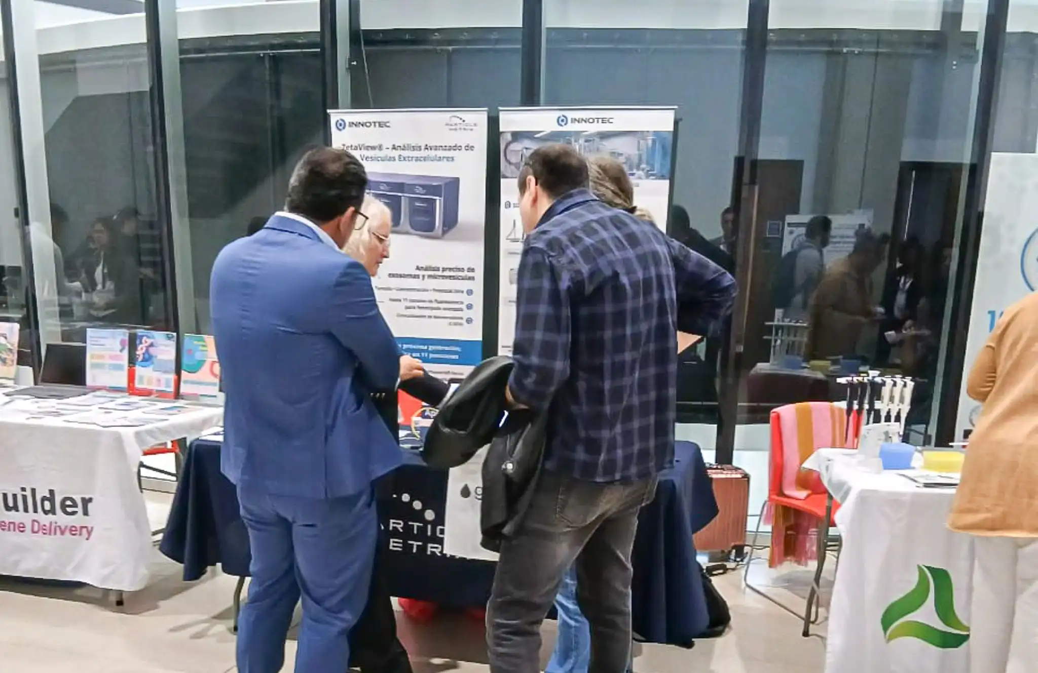
Innotec participated in the IV Workshop as a sponsor, alongside
Particle Metrix
. The manufacturer had also sponsored the 2024 edition, held at UBA.
Rosa Elena Jerlerud Pérez, International Sales Manager at Particle Metrix, gave the talk
ZetaView Evolution: Insights into Bio-nanoparticle Characterization Using NTA
. The presentation covered the characterization of bionanoparticles by nanoparticle tracking analysis, including size, concentration, zeta potential, and identification of subpopulations by fluorescence.
The discussion connected with a need present in several sessions. Studies with placental EVs, bacterial vesicles, and preparations intended for therapeutic applications depend on measurements capable of describing the particles analyzed and on methods suited to the experimental question. No single technique resolves all questions of identity, purity, and function.
What this edition signals
GAVE 2026 presented a field occupied with increasingly specific questions. Researchers tested effects on cells and tissues, evaluated vaccine candidates, modified vesicles to carry cargo, and examined how production and isolation conditions alter the material obtained.
This progress makes characterization more demanding. To compare results across groups and interpret a functional effect, one must know which particles were measured, what may have been co-isolated, and which properties the chosen method preserved. It was this dialogue between biological discovery and methodological rigor that gave unity to such a diverse program.