

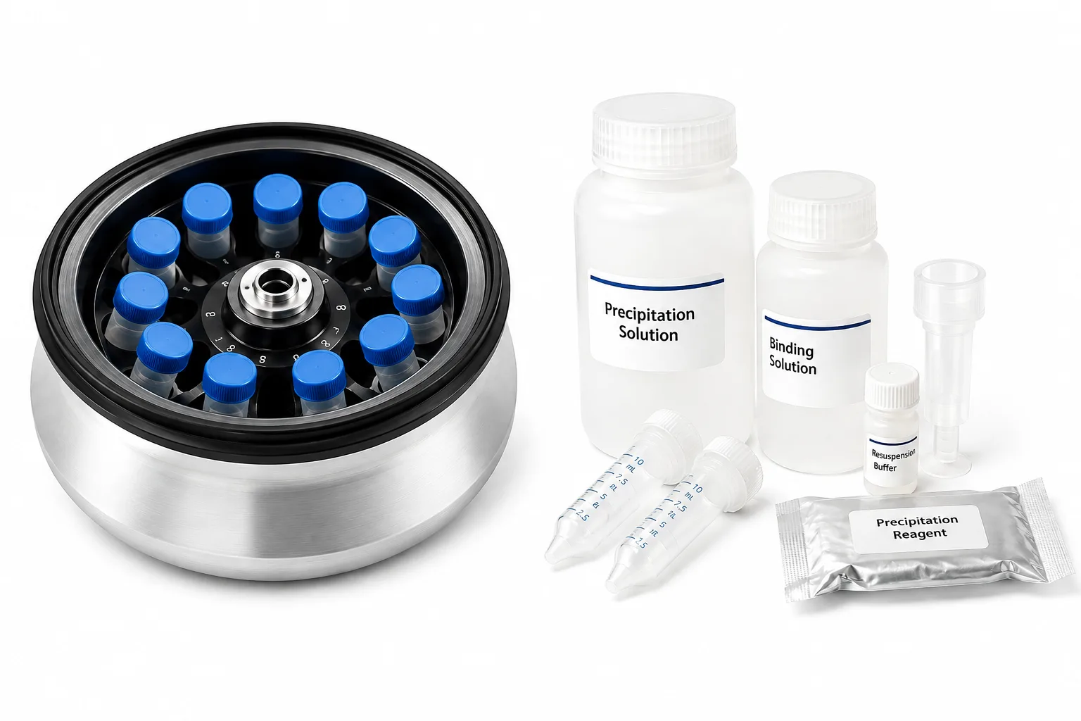
The main exosome extraction techniques are differential ultracentrifugation and precipitation using commercial kits, and a comparative study published in the scientific journal PLOS ONE demonstrated that the three tested kits (miRCURY, ExoQuick, and TEIR) yielded 80 to 300 times more particles than ultracentrifugation, while maintaining equivalent quality.
In this study, the ZetaView instrument, based on nanoparticle tracking analysis ( NTA ), was the central tool used to measure the size, concentration, and zeta potential of the isolated exosomes.
Interest in exosome extraction techniques has grown rapidly over the past decade, driven by the role of these vesicles as potential disease biomarkers and microRNA carriers.
Conducted by researchers from Augusta University in partnership with Duke University , the study compared four isolation methods using different volumes of human serum: differential ultracentrifugation and three commercial precipitation-based kits. Very small volumes were included to simulate the scarcity commonly found in real clinical samples.
The following are the main topics covered in this content:
- Why isolating exosomes is a technical and scientific challenge;
- The four extraction methods compared in the study;
- The role of ZetaView and nanoparticle tracking analysis;
- Complementary characterization methods;
- Main results regarding yield, size, and zeta potential;
- What these findings mean in laboratory practice.
Why Isolating Exosomes Is a Challenge
Exosomes are spherical extracellular vesicles , ranging from 40 to 150 nanometers in size, derived from the endosomal compartment of cells . They carry genomic DNA, RNA, proteins, and lipids, acting as mediators of intercellular communication in multicellular organisms.
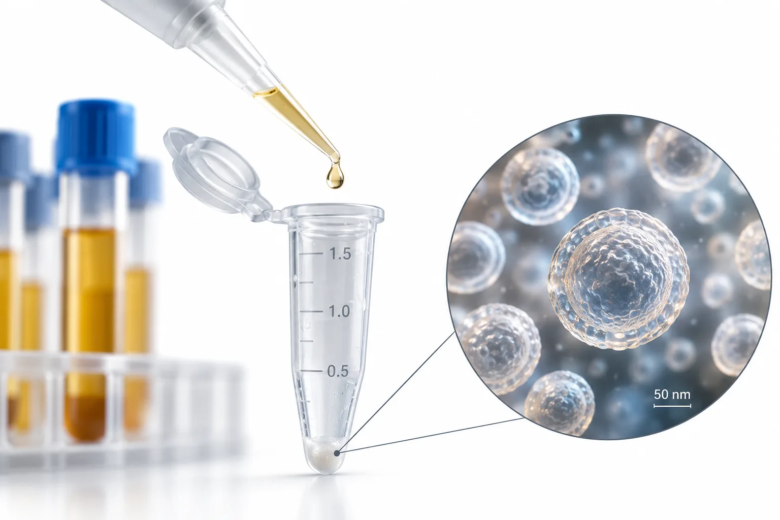
Precisely because they carry microRNA and other signaling molecules, exosomes have gained prominence as candidates for disease biomarkers and potential drug delivery systems. The problem is that isolating them with reliable quality and sufficient concentration is still considered one of the great scientific challenges in the field.
Identifying the ideal isolation technique is essential for any advance in the discovery of new exosome-based biomarkers.
The difficulty lies in separating exosomes from other serum components, such as protein aggregates and lipoproteins, without altering the physical properties or the content of the vesicles.
It is in this context that the choice of extraction method becomes decisive for the validity of the results.
The Four Exosome Extraction Methods Compared
The study compared four isolation methods head-to-head: differential ultracentrifugation and three commercial kits, miRCURY, ExoQuick, and TEIR. Although there are four methods compared, they represent two main isolation approaches: ultracentrifugation and precipitation. The comparison was designed to evaluate the traditional method against different commercial alternatives.
Differential Ultracentrifugation (UC)
Differential ultracentrifugation is the traditional method, adopted for decades as the gold standard for isolating exosomes from biological fluids. The technique separates particles by applying very high centrifugal forces in successive cycles. Although reliable, it is labor-intensive, time-consuming, sensitive to the operator's technique, and requires an ultracentrifuge—an expensive piece of equipment that is not always available.
Commercial Precipitation Kits
The three tested kits, miRCURY (Exiqon), ExoQuick (System Biosciences), and TEIR / Total Exosome Isolation Reagent (Life Technologies), are based on volume-exclusion precipitation, typically using polyethylene glycol (PEG). Therefore, they represent three distinct commercial methods that use the same general isolation approach via precipitation. Compared to ultracentrifugation, they are faster, less technique-sensitive, more compatible with limited sample volumes, and require no special equipment.
How the Study Designed the Comparison
To ensure robustness, the researchers extracted exosomes from six different volumes of pooled human serum (5 mL, 1 mL, 500 μl, 250 μl, 100 μl, and 50 μl) and confirmed the results in six individual donor samples. The smaller volumes were intentionally included to simulate the limited availability of heterogeneous biological samples in clinical studies.
The Role of ZetaView in Exosome Analysis
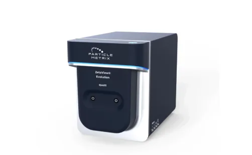
The ZetaView (Particle Metrix) was the central physical characterization instrument in the study. It uses the nanoparticle tracking analysis technique, known by the acronym NTA (from Nanoparticle Tracking Analysis ), which has been used since 2006 as a reliable method for measuring the size and concentration of nanoparticles, including exosomes.
In practice, the equipment tracks the Brownian motion of individual nanoparticles using a video microscope with laser scattering. From this tracking, the software automatically calculates the properties of each sample. The ZetaView is capable of characterizing particles between approximately 10 and 2000 nanometers.
The Three Parameters Measured by ZetaView
The relevance of ZetaView in the study lies in consolidating three essential measurements into a single instrument:
- Size: the diameter of the most abundant particles, used to confirm they are within the expected exosome range (40–150 nm);
- Concentration: the total number of isolated particles, converted to absolute value from the resuspension volume and dilution factors;
- Zeta potential: the electrical potential difference between the fixed layer of the charged particle and the ions in solution, measured in millivolts and used as an indicator of stability.
The greater the magnitude of the zeta potential, the greater the repulsion between particles in solution, suggesting a lower probability of agglomeration or sedimentation. This parameter is, therefore, a direct indicator of the stability of the isolated samples.
According to the authors, this was the first study to integrate zeta potential and absolute microRNA quantification as comparison parameters between isolation methods.
Methods That Complemented Characterization
Although ZetaView was the central axis, the study combined several complementary techniques to validate the identity and quality of the isolated exosomes. This multi-pronged approach was one of the study's differentiators.
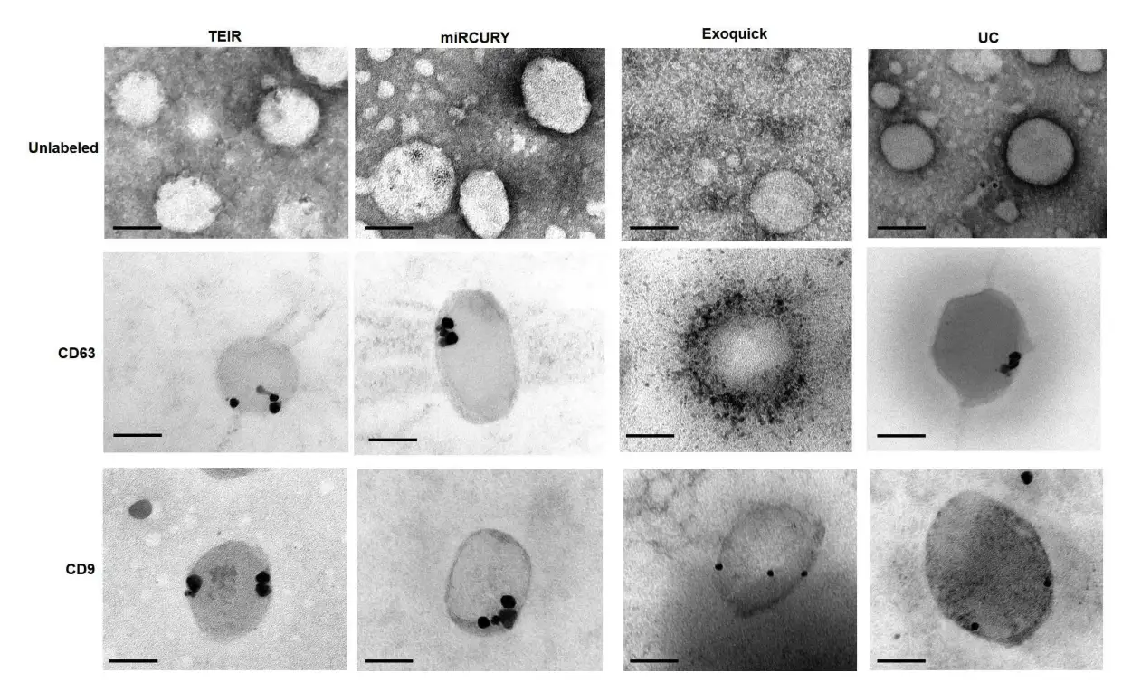
- Transmission electron microscopy (TEM): confirmed the classical morphology of exosomes and, through immunogold labeling, the presence of CD63 and CD9 markers;
- Western blot: detected the expression of CD63 and CD9 proteins in all isolation methods;
- Agilent Bioanalyzer: evaluated the quality and quantity of exosomal RNA;
- ddPCR (droplet digital PCR): absolutely quantified two known microRNAs, miR-16 and miR-451.
Main Results of the Study
The combination of analyses generated a consistent set of results regarding the performance of each isolation method. The findings clarify both the yield and the quality of the isolated particles.
Yield: Kits Surpassed Ultracentrifugation
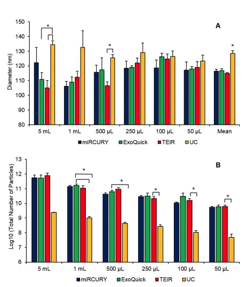
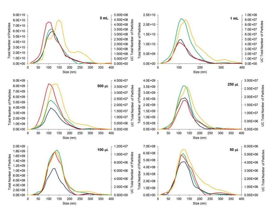
The most significant finding was the difference in yield. The three commercial kits produced a significantly higher number of exosomes than ultracentrifugation in almost all serum volumes. UC isolated approximately 79 to 126 times fewer particles than the kits in volumes from 50 μl to 1 mL.
At the 5 mL volume, the difference reached approximately 299 times, although it did not reach statistical significance due to the large variation between replicates. Furthermore, a linear correlation was observed between serum volume and the number of isolated particles in all methods.
Particle Size and Morphology
All methods isolated particles within the expected exosome range (40–150 nm). In pooled serum, however, ultracentrifugation produced particles with a significantly larger diameter than the kits, an effect attributed to possible fusion of particles with contaminants under the high centrifugation speeds.
Zeta Potential and Stability
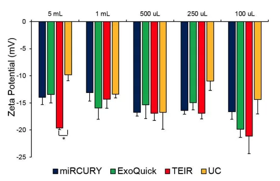
All zeta potential measurements were negative, ranging from -9.8 to -22.6 mV at 23 °C. These values indicate that the isolated exosomes are negatively charged particles, in agreement with previous studies. The relatively low magnitude of the zeta potential points to some instability in solution.
The low magnitude of the zeta potential reinforces the need for caution in handling and storing exosomes, which should be kept at -80 °C.
Quality and Quantity of Exosomal RNA
All extracted RNA showed good quality, with a single peak between 25 and 200 nucleotides and no signs of 18S or 28S ribosomal peaks. Interestingly, although the kits isolated many more particles, this did not translate into a higher amount of RNA: the methods yielded similar amounts of RNA in most volumes.
In ddPCR quantification, all methods detected the microRNAs miR-16 and miR-451, with a positive linear correlation between serum volume and microRNA concentration. The observed variability reinforces the importance of maintaining the same isolation method throughout a study.
What These Findings Mean in Practice
The set of results supports a clear conclusion with direct laboratory application. The three commercial kits, miRCURY, ExoQuick, and TEIR, proved to be suitable alternatives to ultracentrifugation, even with limited amounts of biological material.
In practice, this means that laboratories without access to an ultracentrifuge, or those working with scarce clinical samples, can use the kits without compromising the quality of the isolated material. The main caveat is the possible contamination by lipoproteins, a limitation common to practically all precipitation-based methods.
To overcome this limitation, the authors recommend the consistent and repeated use of the same method within a group of investigated samples, to ensure valid comparisons between them.
Limitations to Consider
The study itself acknowledges limitations in its scope. The comparison did not cover all commercially available reagents, focusing on three of the most used kits. Additionally, only human serum samples were tested, making it necessary to replicate the analysis in other fluids, such as urine, aqueous humor, and cerebrospinal fluid.
Finally, the biological activity of the extracted exosomes was not evaluated, a relevant aspect to confirm the clinical relevance of the vesicles, which could be explored in future studies.
Frequently Asked Questions About Exosome Extraction
What is an exosome?
It is an extracellular vesicle of 40 to 150 nanometers, derived from the endosomal compartment of cells, that carries RNA, DNA, proteins, and lipids and acts in intercellular communication.
What are the main exosome extraction techniques?
Among the approaches evaluated in the study are differential ultracentrifugation and precipitation using commercial kits. Four methods were compared: ultracentrifugation, miRCURY, ExoQuick, and TEIR.
What does ZetaView do in an exosome study?
ZetaView performs nanoparticle tracking analysis (NTA), measuring the size, concentration, and zeta potential of the isolated particles.
Which extraction method yields more exosomes?
In the study, the three commercial kits yielded 80 to 300 times more particles than ultracentrifugation in most serum volumes.
What is zeta potential?
It is the electrical potential difference between the fixed layer of a charged particle and the ions in solution, measured in millivolts. It serves as an indicator of particle stability.
Why should exosomes be stored at -80 °C?
Because the low magnitude of the zeta potential indicates instability in solution, requiring caution in handling and adequate storage temperatures to preserve the vesicles.
Is ultracentrifugation still useful for isolating exosomes?
Yes, it remains a reference method, but it is more labor-intensive, time-consuming, and requires expensive equipment. Kits are viable alternatives, especially when there is little sample available.
What is ddPCR in exosome analysis?
It is droplet digital PCR, a technique used to absolutely quantify specific microRNAs, such as miR-16 and miR-451, present in isolated exosomes.
Related Reading
- Extracellular Vesicles
- Nanoparticle Tracking Analysis (NTA)
- MicroRNA as a Biomarker
Source: DOI:10.1371/journal.pone.0170628